Mohammedelmoez E. A. Mokhtar*, Nadia O Elatta1, Wadah Ali2, Amgad Kh O Nasr3
*Forensic Evidence Administration, Sudan Police Force HQ, Ministry Of Interior
1 National Ribat University –Sudan, Academic Affairs
2 Golf Medical University (GMU), UAE
3 Al-Neelain University – Sudan, Medical Physics Department
*Correspondence e mail: mmmoez@gmail.com, mmmoez@yahoo.com
HNSJ, 2022, 3(10); https://doi.org/10.53796/hnsj3107
Published at 01/10/2022 Accepted at 11/09/2022
Abstract
The radionuclide activities and the fraction of injected activity in the kidneys and bladder have been measured experimentally. The aim of study is to applying the Scintillation Camera Planar Imaging Techniques by using formula derived from the formalism of the Medical Internal Radiation Dosimetry committee, practical data collected from : Radiation and Isotope Center of Khartoum, Nuclear Medicine and Oncology Center, Shendi University, Department Of Nuclear Medicine National Cancer Institute, University of Gazira Wad Medani, Department Of Nuclear Medicine, Royal care International Hospital, Al-Nilain Diagnostic Center- Khartoum. using MedisoInterViewXP® software, The sample of study were randomly selected from available data in sites, it contains 265 patients images referred to Gamma Camera Unit undergoing of bone scan 3 h after injection of methylene diphosphonate (MDP). The measured radionuclide activities were compared with theoretical radionuclide activities values. The radionuclide activities for the kidneys and bladder were found in bone scan 3 h post injection average activity of 20.4 mCi of MDP (0.052 and 0.087mCi) respectively. The study also found that (1.052%) of the injected dose remained in the kidneys, and (1.76%) of the injected dose remained in the bladder three hours post injection, which reflects that low radiation dose was received by the kidneys and bladder during bone scan using 99mTc-MDP. Measured radionuclide activities for the kidneys and bladder were both minimal in the experimental case comparative to the theoretical. The fraction of injected activity in kidneys and bladder were low. The results of the study showed that methods used in the study for Organs dose measurements is in good agreement with the data of MIRDose software, and it is possible to use the obtained method of the present study by a clinician.
Key Words: Radionuclide activities, Nuclear medicine, Internal dosimetry, Radiopharmaceuticals
Introduction/Background:
Nuclear medicine is a medical specialty using radioisotopes as tracers to diagnose diseases or for therapy. These tracers are usually attached to chemical compounds that are attracted to organs of interest such as bones or thyroid gland [1]. The field is divided into diagnosis and therapy. Diagnostic Nuclear medicine is broadly divided into diagnostic imaging, in which biological functions are evaluated based on images obtained by external counting of radionuclide-labeled agents, and in vitro testing, in which trace substances in biological samples such as blood and urine collected from living subjects are measured by radioimmunoassay techniques. Tests using nuclear medicine techniques are more sensitive and specific for disease detection than most tests because they identify abnormalities very early in the progression of a disease [2]. Special electronic instruments such as scintillation or a gamma camera, which displays these emissions into images, can detect these emissions [3]. The role of internal dosimetry in diagnostic nuclear medicine is thus to provide the basis for stochastic risk quantification. Once this risk is quantified, it may be used to optimize the amount of administered activity in order to maximize image quality while minimizing patient risk. This optimization is considered, and always evaluated for any imaging procedure [4]. A bone Scan or scintigraphy is a nuclear medicine test to find certain abnormalities in bone; it is primarily used to help diagnose a number of conditions relating to bones [5]; most bone disease needs high quality anatomical imaging, involving radiography, CT or MRI. Nuclear Medicine studies are best undertaken when physiological information is required. Whole-body bone scintigraphy produces planar images of the skeleton.
All nuclear medicine investigations cause exposure to ionizing radiation of the whole body. Using properly conducted diagnostic nuclear medicine procedures the mean absorbed doses to various organs and tissues are, in general, low. The risk of health effects due to radiation is, however, not negligible, since it is stochastic. Therefore radiation protection is always essential [6].
The main objective of this study is to measure the organs dose during the Nuclear Medicine scans in Sudan, specifically kidneys and bladder during Bone scan, using Scintillation Camera Planar Imaging Techniques. Most scintillation camera systems used in clinical nuclear medicine are computer-based and provide software for generation of ROIs and statistical analysis [7], then compare it with the theoretical values published in scientific paper by Luc T. Bambara el at, which determined the activity in kidneys and bladder from time-activity curves stimulated by using Compartmental Modeling published in Medical Internal Radiation Dosimetry committee reports for radiopharmaceuticals [8]
Materials and Methods:
The data of this study was collected from the five main centers of Nuclear Medicine in Sudan: Radiation and Isotope Center of Khartoum, Nuclear Medicine and Oncology Center, Shendi University, Department Of Nuclear Medicine National Cancer Institute, University of Gazira Wad Medani, Department Of Nuclear Medicine, Royal care International Hospital, Al-Nilain Diagnostic Center- Khartoum. The sample of study were randomly selected from available data in sites, it contain 265 patients, undergoing of bone scan, 3 hrs post injection of average activity of 20.4mCi of methylene diphosphonate (MDP). Single photon emission computed tomography (SPECT) system (Mediso manufacture) was used for performing bone scans on patients as employed in studies, the data was obtain by using Scintillation Camera Planar Imaging Techniques, and calculated radionuclide activity for the kidneys and bladder by using formula derived from the formalism of the Medical Internal Radiation Dosimetry committee (MIRD), the biodata for patients was obtained, Quantitative analyses were performed on images acquired by drawing region of interests (ROIs) by the system’s software application Mediso / Medical Imaging systems –InterView XP, for the kidneys and bladder, also for whole body, anterior and posterior, and tabulating counts of radionuclide activity, the average count were calculated from the conjugate view counting by using geometric mean count equation : GMC = where and are the anterior and posterior counts respectively, then the count of right Kidney and left kidney and bladder were calculated and the average kidney count was obtained, the fraction of counts in kidneys and bladder were calculated from whole body count, and then the radionuclide activity in kidneys and bladder were estimated by looking for an average of injected dose with the whole body counts and compare the fraction of activity in kidneys and bladder with that ratio, and the counts were converted into radionuclide activity (mCi).
Then the measured radionuclide activities were compared with the theoretical radionuclide activities value.
The results acquired were compared by t-test (independents samples test) with data of the theoretical radionuclide activities. The whole results were considered statistically significant difference at p <0.001.
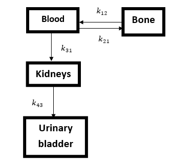
Fig (1) Biokinetic Model for () MDP for Bone scan

Compartment 1 represents blood and extracellular fluid (ECF); compartment 2, bone; compartment 3, kidneys; and compartment 4, urine.
Results and Discussion:
The values of measured and theoretical radionuclide activities:
The radionuclide activity of kidneys and Bladder during Bone scan (mCi):
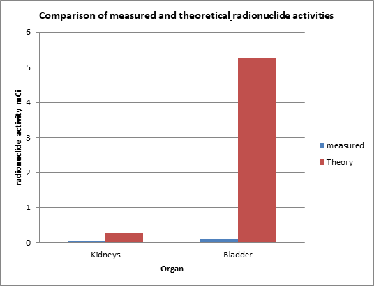
| Organ | measured | Theory |
| Kidneys | 0.052 | 0.270 |
| Bladder | 0.087 | 5.276 |
The comparison of measured and theoretical radionuclide activities:

The values of the fraction measured and theoretical radionuclide activities:
The fraction radionuclide activity of kidneys and Bladder during Bone scan %:
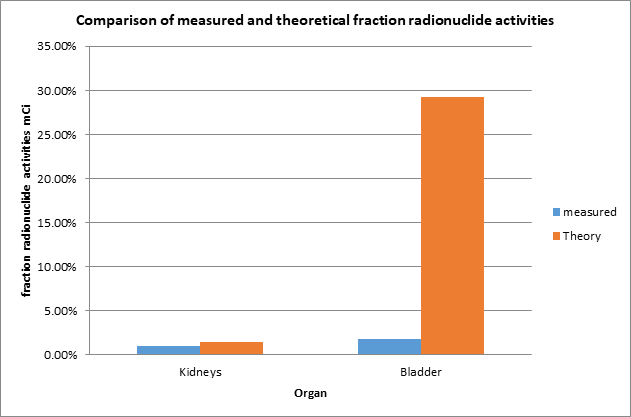
| Organ | measured | Theory |
| Kidneys | 1.05% | 1.5% |
| Bladder | 1.76% | 29.31% |
The comparison of measured and theoretical fraction radionuclide activities:

Radionuclide activity in kidneys and bladder:
The study found that the experimental measured radionuclide activity in kidneys and bladder in bone scan for 265 patients 3 h post injection of average activity 20.4 mCi of MDP are (0.052 and 0.087mCi) respectively. The study also found that 1.05% of the injected dose remained in the kidneys, and 1.76 % of the injected dose remained in the bladder in bone scan for 265 patients 3 h post injection of average activity20.4 mCi, which reflects that low radiation dose was received by the kidneys and bladder during bone scan using 99mTc-MDP.
The study found that the measured radionuclide activity in kidneys is (0.052 mCi) is less than the theoretical value which is (0.27 mCi) and the measured radionuclide activity bladder radionuclide activity (0.087 mCi) is less than the theoretically estimated values which is (5.276 mCi) during Bone scan. Which the values of radionuclide activities are depending on the injected dose.
The study found that the measured fraction of radionuclide activity in kidneys is (1.05%) is less than the theoretical value which is (1.5%). And the measured fraction radionuclide activity bladder radionuclide activity (1.76%) is less than the theoretically estimated values which is (29.31%) during Bone scan. and that because in the practice the patients ordered to drink a lot of water and ordered to urinate and that minimize the activity in bladder which “the residence time for bladder contents was calculated by solving the differential equation that describes the change in activity in Compartment 4 (urine) and using a model in which the bladder was emptied at 2 h after administration of the radiopharmaceutical and then at 4.8-h intervals, integrating the activity” as a noted in [9].
The results acquired were compared by t-test (independents samples test) with data of the theoretical radionuclide activities. The whole results were considered statistically significant difference at p <0.001.
Conclusions:
The results of this study showed that methods used in the study for Organs dose measurements is in good agreement with the data of MIRDose software, and it is possible to use the obtained method of the present study by a clinician,
Also findings that may be useful to estimate the amount of activity that can be administered to the patients and serve as a way of comparing the risk to the benefit value of these nuclear medical procedures with the other modalities of diagnostic procedures especially for the affected organs to keep as low as possible.
Acknowledgment:
I would like to give my thanks to administration and staff of the five centers of Nuclear Medicine in Sudan, for their helpful stand in data collection, Radiation and Isotope Center of Khartoum, Nuclear Medicine and Oncology Center, Shendi University, Department Of Nuclear Medicine National Cancer Institute, University of Gazira Wad Medani, Department Of Nuclear Medicine, Royal care International Hospital, Al-Nilain Diagnostic Center- Khartoum.
Reference:
- Bolch, W, & Fahey, F. “The management of imaging procedure dose. Nuclear Medicine, 2” available at https//www.aapm.org/meetings/2013AM/PRAbs.asp? Retrieved 14.8.2016.
- Michael Lassman, Markus Luster, HeribertHanscheid, and ChristophReiners. “Impact of Diagnostic Activities on the Biokinetics of Thyroid Remnants”. The Journal of nuclear medicine, J. Nucl. Med; 45: 619-625. 2004
- Michael G. Stabin, “The Importance of Patient-Specific Dose Calculations in Nuclear Medicine”. Nuclear Engineering and Technology. Vol. 40, No. 7. 2008
- IAEA, (2011), “Report of a consultancy meeting”, International Atomic Energy Agency, Vienna: IAEA. held 26-28 September 2011.
- DaryoushShahbazi‑Gahrouei, Mohsen Cheki1, MasoudMoslehi, “Estimation of Organ Absorbed Doses in Patients from 99mTc‑diphosphonate Using the Data of MIRDose Software”, Short Report , Journal of Medical Signals & Sensors, Vol 2 | Issue 4 | Oct-Dec 2012, page: 231
- Sigrid Leide Svegborn, “Radiation Exposure of Patient in Diagnostic Nuclear Medicine”, Copyright@ 1999 Sigrid Leide Svegborn ISBN 91-628-3491-6 Printed in Sweden by KF-Sigma, Lund 1999
- MIRD16, Jeifry A. Siegel, el at, “Techniques for Quantitative Radiopharmaceutical Biodistribution Data Acquisition and Analysis for Use in Human Radiation Dose Estimates”, THE JOURNALOFNUCLEARMEDICINE, Vol. 40. No. 2 (Suppl). 1999, page 415
- Luc T. Bambara, Augustine K. Kyere, Francis Hasford, “Estimation of kidney and bladder radionuclide activity for patients undergoing bone scan”, Journal of Radiation Research and Applied Sciences. 2015, page 320
- MIRD13, David A. Weber, el at, “Radiation Absorbed Dose from Technetium-99m-Labeled Bone Imaging Agents”, THE JOURNALOFNUCLEARMEDICINE, Volume 30 Number 6. 1989. page 1119
