

Evaluation of Marginal Accuracy of Two Ceramic Restorations Using Different Impression Materials
Ezzedden Alhur1
1 Fixed Prosthodontic Department, Faculty of Dentistry, Islamic University (Libya).
Email: dr.ezzofree77@gmail.com
DOI: https://doi.org/10.53796/hnsj62/15
Arabic Scientific Research Identifier: https://arsri.org/10000/62/15
Volume (6) Issue (2). Pages: 174 - 181
Received at: 2025-01-04 | Accepted at: 2025-01-15 | Published at: 2025-02-01
Abstract: A variety of impression materials are available on the market, necessitating numerous studies to assess their accuracy and its impact on the marginal fit of final restorations. This research indicates no significant differences in accuracy among the materials investigated.
Keywords: Marginal Accuracy, Ceramic Restorations, Impression Materials.
Introduction
Impression materials are utilized to capture intraoral structures. Ideally, these materials should exhibit high accuracy and minimal distortion, enabling the recording of fine details without requiring additional impressions. They are designed to be soft, elastic, and tear-resistant, allowing for easy removal from undercuts.
Poorly fitting prosthetic restorations can adversely affect periodontal health and occlusion. The design of tooth preparations is a crucial aspect of tooth reconstruction. The geometric features employed in full-coverage restorations are often based on clinician experience and personal preference.
The dimensional stability of impression materials can influence the fit and retention of restorations, thereby impacting the success of indirect restorative procedures. Factors such as humidity, the time elapsed from mixing to pouring, and the thickness of the material layer in the tray can affect the dimensional behavior of these materials. Additionally, temperature changes from the oral cavity to the external environment may cause impression materials to contract, which is related to their linear thermal expansion coefficient.
With new generations of impression materials continuously emerging in the dental market, further studies are essential to investigate their properties and validate their applications. This study aims to determine which impression materials can ensure accurate seating of final restorations.
Materials and Methods
This study evaluates the accuracy of three different impression materials in conjunction with two types of ceramic materials: zirconia and lithium disilicate, utilizing machinable (CAD/CAM) technology (CEREC in Lab). The impression materials used in this research include
Methodology
Sample Grouping
The impression materials were categorized into the following groups:
- Group I: Laminated hydrocolloid technique
- Group II: Addition silicone impression materials
- Group III: Polyether impression materials
Each group was further divided into two subgroups based on the ceramic material used:
- Subgroup A: Zirconia (15 samples)
- From each impression material, 5 non-anatomical CAD/CAM ceramic crowns were fabricated.
- Subgroup B: Lithium disilicate (15 samples)
- Similarly, for each impression material, 5 non-anatomical CAD/CAM ceramic crowns were created.
Die Fabrication
To ensure standardization, a custom master die was constructed to represent a full all-ceramic crown preparation, which facilitated standardized impression making. A perforated custom tray was designed to be positioned consistently on the master die for each impression.
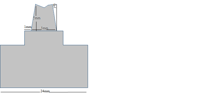
The metal master die was created using a milling machine, designed to mimic a prepared mandibular second premolar. It measured 5 mm in height and 5 mm in width at the base, with an occlusal taper of 6°. The occlusal surface featured two sloping surfaces, one of which was slightly beveled. The preparation ended with a rounded shoulder finish line that was 1 mm wide. This design was selected to prevent rotation of the crowns on the die and to allow for reproducible placement. Four equidistant marks were engraved on the die to orient the stereomicroscope, and the die had a 24 mm diameter base for proper handling.
Custom-Made Perforated Tray
Each custom-made perforated tray was cylindrical, featuring an inner diameter of 7 mm to fit the base of the master die and an inner height of 7 mm. These trays were designed to hold the impression materials (see Fig. 2).
Impression Making
A single impression was taken for each material on the same metal die using the perforated trays. All fabrication processes were conducted according to the manufacturer’s guidelines for each material tested.
- Laminated Hydrocolloid Technique:
Impressions were obtained using agar alginate impression material. The agar was heated in boiling water for approximately six minutes and then maintained at 65°C for at least 10 minutes before being syringed around the preparations. A mixture of alginate was prepared per the manufacturer’s instructions and placed in the custom-made perforated tray, which was then seated over the agar material. Once the alginate set, the combined impression was removed (see Fig. 3).
- Polyvinyl Siloxane Impression:
The single-step impression technique was employed, where heavy-bodied material was injected into the custom-made perforated trays. Concurrently, light-bodied material was injected onto the die. The impression tray was centered over the die and seated with finger pressure to ensure proper positioning and allow excess material to escape. After complete setting, the tray was removed sharply in a parallel motion to the long axis of the die (see Fig. 4).
- Polyether Impression Materials:
Equal portions of impression base paste and catalyst were placed on a paper pad and mixed with a metal spatula in circular motions to achieve a homogeneous mixture. This mixture was then loaded onto the perforated tray and applied to the die. The impression tray was centered over the die, and after setting, it was removed parallel to the long axis of the die.
Crown Construction
Cylindrical crowns, measuring 6 mm in diameter, 7 mm in height, with a 2 mm occlusal thickness and a 1 mm margin thickness, were constructed for each material according to the manufacturer’s instructions (see Fig. 6). All tested crowns were individually seated on the stainless steel die and examined for vertical marginal fit relative to the die.
Testing Procedures
Cervical Marginal Accuracy
To assess the cervical marginal accuracy of the crowns, they were seated on a stainless-steel die and secured in place for examination using a stereomicroscope. Digital images of the margins for each crown were captured with a camera attached to the stereomicroscope, using a fixed magnification of 40X (see Fig. 2a, b).
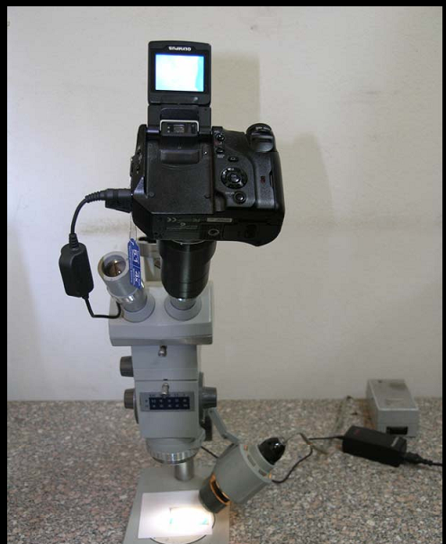
Equipment Used:
Carl Zeiss Stereomicroscope, Germany Olympus Camedia C-5060 Digital Camera, Japan
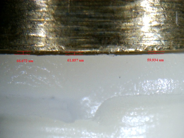
Morphometric measurements were conducted on an IBM-compatible personal computer. The image analysis software was calibrated prior to use, allowing for precise measurements of the vertical gap distance for each captured image. Measurements were taken at six equidistant landmarks along the cervical circumference of each crown: mesio-buccal, mid-buccal, disto-buccal, mesio-lingual, mid-lingual, and disto-lingual line angles. Each measurement point was recorded five times to ensure accuracy. The collected data were then tabulated and subjected to statistical analysis.
Vertical Marginal Gap
The mean values and standard deviations of the vertical marginal gap (in micrometers) as a function of the ceramic and impression materials are summarized in Table 5 and illustrated graphically in Figure 21. The results indicated that e.max CAD restorations exhibited the highest marginal gap (62.19 µm) with polyether impression material, while the lowest marginal gap was observed with the laminated impression technique
Table (5 ) Vertical marginal gap results (Mean values± SDs) as function of ceramic and impression materials
|
Variables |
Ceramics |
Statistics |
||||||
|
Zr |
e.max |
t-test |
||||||
|
Mean |
SD |
Mean |
SD |
P value |
||||
|
Impression materials |
Laminate |
38.41Aa |
19.87 |
36.12Ca |
9.44 |
0.5635 ns |
||
|
Addition silicon |
41.83Aa |
10.67 |
46.57Ba |
11.04 |
0.0718 ns |
|||
|
Polyether |
42.34Ab |
10.14 |
62.19Aa |
18.99 |
<0.0001* |
|||
|
Statistics |
(P-value) |
0.4795 ns |
<0.0001* |
|||||
Superscripts indicate statistically significant differences among impression materials (p < 0.05), while subscripts indicate differences among ceramic materials (p < 0.05).
significant (p < 0.05) ns; non-significant (p>0.05)
Figure (3 ) Histogram of vertical marginal gap mean values as function of ceramic and impression materials
Table (6 ) Two factorial analysis of variance ANOVA test of significance comparing variables affecting vertical marginal gap mean values
|
Source of Variation |
Df |
SS |
MS |
F |
P value |
|
Ceramics |
1 |
3978.95335 |
3978.95335 |
13.2411 |
0.0003* |
|
Impression materials |
2 |
10817.70351 |
5408.85176 |
17.6926 |
<0.0001* |
|
Interaction |
2 |
6141.01819 |
3070.50909 |
10.0438 |
<0.0001* |
Effect of ceramic
Regardless of the impression materials used, the e.max group exhibited a statistically significant higher vertical marginal gap mean value (48.29 ± 9.26 µm) compared to the Zirconia (Zr) group (40.86 ± 1.63 µm), as indicated by two-way ANOVA followed by pairwise Tukey’s post-hoc tests
Table ( 7) Comparison between total vertical marginal gap results (Mean values± SDs) as function of ceramic
|
Variable |
Mean |
SD |
Tukey’s rank |
Statistics (P value) |
|
|
Ceramic |
Zr |
40.86 |
1.63 |
B |
0.0003* |
|
e.max |
48.29 |
9.26 |
A |
||
Different letter in the same column indicating statistically significant difference (p < 0.05)
*; significant (p < 0.05) ns; non-significant (p>0.05)
Figure (4) A column chart of total vertical marginal gap mean values as function of ceramics
Discussion
The accurate transfer of a patient’s hard and soft tissue to the dental laboratory is crucial for fabricating both fixed and removable restorations. Producing a definitive impression is a vital step in creating restorations that are biologically, mechanically, functionally, and esthetically acceptable (84).
One of the key factors in the selection of impression materials for fixed prosthodontics is the ability to achieve a smooth surface and precise details on stone models (59). This precision is essential for ensuring that the final restoration fits properly and functions effectively within the oral environment. Inadequate impression accuracy can lead to complications, including improper occlusion, increased wear, and patient discomfort.
The study also examined the laminated hydrocolloid technique in comparison to other popular impression materials, such as vinyl polysiloxane and polyether. Recently, a combination of reversible (agar) and irreversible hydrocolloid (alginate) impression systems has been introduced into dental practice. This method, known as the “laminated hydrocolloid technique,” involves injecting reversible hydrocolloid onto the prepared tooth, followed by positioning a custom tray loaded with irreversible hydrocolloid over it.
During this process, the alginate sets through a chemical reaction while the agar gelates due to contact with the cooler alginate, rather than being cooled by water in the tray (94). This innovative approach aims to enhance the accuracy and detail of the impression, potentially addressing some limitations associated with traditional materials. The findings of this study will contribute to a deeper understanding of the effectiveness of various impression techniques in clinical practice, ultimately improving prosthodontic outcomes.
Summary & Conclusion
This in vitro study investigated the marginal accuracy of various impression materials using two types of ceramic materials through machinable Computer-Aided Design/Computer-Aided Manufacturing (CAD/CAM) techniques. The results of the study revealed the following:
- The laminated hydrocolloid agar-alginate impression technique demonstrated a significantly lower marginal gap compared to both polyvinyl siloxane and polyether materials.
- For zirconia copings, there was no significant difference in marginal gap between polyether and polyvinyl impression materials.
- In the case of e.max CAD copings, a significant difference in marginal gap was observed between polyether and polyvinyl impression materials.
Within the limitations of this study, it can be concluded that the laminated hydrocolloid agar-alginate impression technique can be utilized as a secondary impression method with high accuracy.
References
1.Kronström MH, Johnson GH, Hompeschc RW (2010), ‘Accuracy of a new ring-opening metathesis elastomeric dental impression material with spray and immersion disinfection’, J Prosthet Dent vol. 103 pp 23-30.
2.Quintas AF, Oliveira F, Bottino MA.: “Vertical marginal discrepancy of ceramic copings with different ceramic materials, finish lines, and luting agents.” J Prosthet Dent. 2004; 92:250-257
3. Shah S, Sundaram G, David B and Sherriff M : The use of a3D laser scanner using software superimposition to assess the accuracy of impression techniques. J. Dent., 2004; 32:653.658.
4. Marcinack CF, Young FA, Draughn RA and FlemmingWR: Linear dimensional changes in elastic impression materials. Dent. Res., 1980; 59:1152.1155.
5. Mehta R, Wadhwa S, Duggal N, Kumar A2, Goel M and Pande S: Influence of Repeat Pours of Addition Silicone Impressions on the Dimensional Accuracy of Casts. J Interdiscipl Med Dent Sci 2014;
6. Chen SY, Liang WM and Chen FN: Factors affecting the accuracy of elastomeric impression materials. J. Dent., 2004; 32:603.609.
7. R.G. CRAIG, Reiviw of dental impression materials Presented at the International State-of-the-Art Conference on Restorative Dental Materials, Adv Dent Res 2(l):51-64, August, 1988
8. Groten MS, Girthofer S, Probster L. Marginal fit consistency of copy-milled allceramic crowns during fabrication by light and scanning electron microscopic analysis in vitro. J Oral Rehabil 1997;24:871-81
9.Gassino G, Barone Monfrin S, Scanu M, Spina G, Preti G. Marginal adaptation of fixed prosthodontics: A new in vitro 360-degree external examination procedure. Int J Prosthodont 2004;17:218-23.
10.Gu XH, Kern M Gu XH, Kern M. Marginal discrepancies and leakage of all-ceramic crowns: influence of luting agents and aging conditions. Int J Prosthodont 2003;16:109-16.
11. Albert FE, El-Mowafy O. M. Marginal adaptation and microleakage of Procera AllCeram crowns with four cements. Int J. Prosthodont 2004;17:529-35.
12. Vahidi F and Egloff E, panno F , Evalution of marginal adaption of all-ceramic crowns and metal ceramic crown . J Prosth dent 63:426,1991
13. Clpe X, James L, Martin F. Bond strength and accuracy of combined reversible-irreversible hydrocolloid impression system. J Prosthet Dent 1992;67(5):621-32.
14. Karatasli O, Kursoglu P, Çapa N, Kazazoglu E. Comparison of the marginal fit of different coping materials and designs produced by computer aided manufacturing systems. Dent Mater J 2011;30(1).
15. Jidige Vamshi Krishna, sumanth Kumar, Ravindra Savadi.: Evolution of metal-free ceramics. J.india prosth.2009;11:72-74
16. Anusavice KJ and Kenneth J: Phillips’ science of dental materials. 11th edition. Elsevier 2003. p. 12.
17. Craig RG: Restorative Dental Materials (ed 11). St Louis,Elsevier, 2001. 48-58.
18.Schneider W.: No compromises the new CEREC MC XL and inLab MC XL milling machines. Int. J. Comput.Dent. 2007;10:119-126
19.Kurbad A.: CEREC goes inLab the metamorphosis of the system.Int. J. Comput. Dent. 2001;4:125-
20. Appleby DC. The combination of hydrocolloid/alginate impression. JADA 1983;106:194-
21. Anusavice KJ: Phillips’ Science of Dental Materials (ed 11). Philadelphia, Saunders, 2003, p. 216.
22.Helmer J.D., Driskell T.D.: Research on bioceramics. Symposium on Use of Ceramics as Surgical Implants. South Carolina, USA: Clemson University; 1969
23. Hembree JH JR and Nunes LJ : effect of moisture on polyether impression materials. J Am Dent Assoc. 1974; 89(5): 1134-6.
24. Klettke T, Kuppermann B, Führer C, and Richter B: Hydrophilicity of Precision Impression Materials During Working Time. 3M ESPE AG, Seefeld, Germany.
25.Green D., Hannink R., Swain M.: Transformation toughening of ceramics. Boca Raton, FL: CRC Press; 1988
26.Hannink R.H.J., Kelly P.M., Muddle B.C.: Transformation toughening in zirconia-containing ceramics. J. Am. Ceram. Soc. 2000;83:461–487
27. Skinner WE, Hoblet N. A study of accuracy hydrocolloid impression. J Prosthet Dent 1956;6(1):80-8
28. Sorensen J, Astandared method for determination of crawon margin fidelity ,J Prosthet Dent 1990;64:18-24.
29, Melean W Wilson D , prosser J . Development and use of water hardening glass ionomer luting cement . J Prosthet Dent 1984;52:175-81
30. Shakila Fatemaa, Sheikh Md. Shahriar Quaderb, Mohammad Shamsuzzamanc,Mirza Md. Arifur Rahmand, Nasima Khane A Comparative Study on Accuracy and Reproducibility of Alginate and Addition Reaction Silicone as an Impression Materials Updat Dent. Coll .j 2013; 3(2):28-33.
31.Felton D, Kanoy B, Bayne S and Wirthman G . effect of in vivo crown margin discrpancies on periodental health . J Prosthet Dent 1991;65:357-64.
32. Hondrum S. O.: A review of the strength properties of dental ceramics. J. prosth. Dent. 1992;67:859-33.Swab J.J.: Low temperature degradation of Y-TZP materials. Journal of Materials Science 1991;26:6706–6714
34.Luthardt R.G., Holzhuter M., Sandkuhl O., Herold V., Schnapp J.D., Kuhlisch E.: Reliability and properties of ground Y-TZP-zirconia ceramics. Journal of Dental Research 2002;81:487–491
35.Kosmac T., Oblak C., Jevnikar P., Funduk N., Marion L.: The effect of surface grinding and sandblasting on flexural strength and reliability of Y-TZP zirconia ceramic. Dental Materials 1999;15:426–433
36. Pera P., Gilodi S., Bassi F., Carossa S.: In-vitro marginal adaptation of alumina porcelain ceramic crowns. J. Prosthet. Dent.1994;72:585-590
37. Wolfart S Wegner SM, AL-Halabi A and Kern M. Clinical evalution of marginal fit of new all ceramic system befor and after cemention. Int J Prosthodont 2003;16:587
38. . Park SH, Lee KB. A comparison of the fidelity between various cores fabricated with CAD/CAM system. J Kor Acad Prosthodont 2008;43(3): 269-78
39. Atousa Azarbal, marginal fit comparison of cad/cam crowns milled from two different materials University of Pittsburgh, 2015;24:871-88
40 . Tinschert J, Natt G, Mautsch W, Spiekermann H. Marginal fit of alumina- and zirconia-based fixed partial dentures produced by a CAD/CAM system. Oper Dent 2001;26:367-74.
41. Beuer F, Aggstaller H, Edelhoff D, Gernet W, Sorensen J. Marginal and internal fits of fixed dental prostheses zirconia retainers. Dent Mater 2009;25:94-102.
42. Donovan TE, and Chee, WW: A review of contemporary impression materials and techniques. Dent. Clin. N. Am., 2004;48:445.470.
43. APPLEBY, D.C.; PAMEIJER, C.H.; and BOFFA, J. (1980): The Combined Reversible Hydrocolloid/Irreversible Hydrocolloid Impression System, / Prosthet Dent 44:27-35.
44. Giordano R: Impression materials: basic properties. GenDent 2000; 48:510-516.
45. Appleby DC, Cohen SR. The combined reversible/irreversible hydrocolloid impression system: Clinical implication. J Prosthet Dent 1981;46(1):48-58
46. Heering HW, Tames MA. Comparison of the dimensional accuracy of a combined reversible/irreversible hydrocolloid impression system with commonly used impression materials. J Prosthet Dent 1984;52(6):795-99.
47. Pace SL. Polyvinyl impression materials vs. alginate impression materials. Contemp Dental Assisting. Feb 2006:20-23.